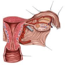
Uterine Arteriovenous Malformations (AVM)
Uterine arteriovenous malformations (AVMs) are rare and complex vascular disorders that can lead to serious health problems in women. These abnormalities occur due to abnormal connections between arteries and veins, which may cause severe bleeding, pain, and other complications. With advances in medical technology, new treatment approaches have been developed — one of the most effective being embolization performed by interventional radiology.
This article explores the details of uterine AVMs, the embolization procedure, its steps, benefits and drawbacks, as well as post-treatment care.
What is a Uterine Arteriovenous Malformation (AVM)?
Definition
An arteriovenous malformation (AVM) is a condition in which arteries connect directly to veins without the normal capillary network in between. This leads to increased pressure in the veins, causing them to enlarge. AVMs can occur anywhere in the body, but when they develop in the uterus, they may result in unique complications.
Causes of AVM
The exact cause of AVM is not fully understood, but several factors may contribute:
- Congenital: Some AVMs are present at birth.
- Trauma or injury: Physical injury to the pelvis or uterus may trigger AVM formation.
- Genetic factors: Family history may increase susceptibility.
Symptoms
Symptoms of uterine AVM may include:
- Severe vaginal bleeding: A hallmark sign of AVM, which can be life-threatening.
- Pelvic pain: Chronic or sudden pain in the pelvic region.
- Frequent urination: Enlarged vessels may press against the bladder.
- Fatigue and weakness: Often due to anemia from recurrent bleeding.
What is Interventional Radiology?
Interventional radiology is a specialized branch of radiology that uses imaging techniques to perform minimally invasive diagnostic and therapeutic procedures. These procedures employ catheters, needles, and other tools to treat conditions with minimal damage to surrounding tissues.
Advantages of Interventional Radiology
- Minimally invasive: No need for open surgery.
- Shorter recovery time: Patients return to normal life more quickly.
- Fewer complications: Lower risk compared to traditional surgery.
- High precision: Imaging guidance ensures accuracy in treatment.
Embolization for Uterine AVM
Definition
Embolization is a non-surgical interventional technique used to block specific blood vessels. It is commonly used to control bleeding, reduce blood flow to tumors, or treat vascular malformations. In uterine AVM, embolization blocks the affected vessels, lowering blood pressure within them and relieving symptoms.
Steps of Embolization
- Patient preparation: Pre-procedure evaluations and detailed explanation to the patient.
- Local anesthesia: The catheter insertion site is numbed.
- Catheter placement: Under imaging guidance (e.g., ultrasound or CT), a catheter is advanced into the targeted blood vessels.
- Injection of embolic agents: Special materials such as polymer particles or coils are injected to block the abnormal vessels.
- Monitoring: The patient is observed after the procedure to detect any complications.
Benefits and Drawbacks of Embolization
Benefits
- Symptom relief: Many patients experience significant improvement.
- Minimally invasive: No need for open surgery and faster recovery.
- Uterus preservation: Unlike hysterectomy, this procedure allows the uterus to be preserved.
Drawbacks
- Possible side effects: Like any medical procedure, risks exist.
- Recurrence: In some cases, AVM symptoms may return.
- Need for follow-up: Regular medical monitoring is required to ensure long-term results.
Potential Complications of Embolization
Although generally safe, embolization may carry some risks:
- Post-procedure pain: Some patients may feel discomfort.
- Infection: Risk at the catheter insertion site.
- Re-bleeding: If embolization does not fully occlude the vessels.
- Damage to nearby tissues: Rare but possible complication.
Post-treatment Care
Following embolization, patients should be monitored to prevent complications. Recommended care includes:
- Adequate rest: Several hours of rest after the procedure.
- Monitoring vital signs: Regular checks of blood pressure and pulse.
- Avoiding strenuous activity: Physical exertion should be limited for a few days.
- Observation of warning signs: Severe pain, fever, or bleeding should be reported immediately.
Conclusion
The treatment of uterine arteriovenous malformations (AVM) with embolization through interventional radiology is an effective and minimally invasive approach for managing vascular abnormalities in the pelvic region. Given its multiple advantages, it is a valuable option for patients who are not candidates for surgery or those seeking non-surgical alternatives.